Watching the inner life of a cell
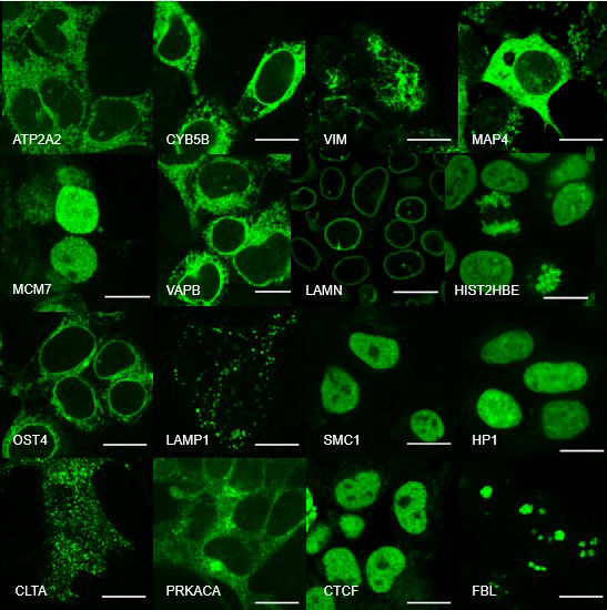
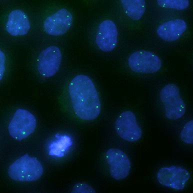
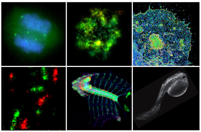
Cellular
processes are orchestrated by a large number of biomolecules in a spatially
and temporally coordinated manner within a tiny volume. To uncover the
underlying organizational principles and their functional relevance, we take
microscopy visualization as the primary approach to systematically map the
spatial localization, temporal dynamics and activity profiles of proteins
and nucleic acids.
We are particularly interested in the following problems:
- Subcellular, high-resolution and dynamic mapping of the entire proteome,
- Physical organization and dynamics of the nucleus and the genome,
- Architecture of large protein complexes such as the ciliary transition zone, and
- Subcellular compartmentalization of signaling molecules, particularly
in the G-protein coupled receptor (GPCR) and receptor tyrosin kinase (RTK) signaling pathway, and how this spatial distribution defines signaling specificity.
In order to study these systems, we are developing the following microscopy technologies:
- Super-resolution and light-sheet microscopes that can visualize subcellular structures at a higher spatial resolution, record long term cell behavior, and track cells in intact animals, and
- New fluorescent probes and gene editing methods for the labeling of
endogenous proteins.
Research updates
![]() |
![]() |
![]() |
![]() |
Images from protein sequences bioRxiv |
The OpenCell Science |
Image denoising by transfer learning Opt. Express |
CryoEM for endogenous complexes bioRxiv |
Lab updates...
We welcome postdoc applicants and new graduate students:
Please email your CV to and arrange to
send three letters of reference if you are interested in becoming a postdoc
in the lab.
For perspective graduate students, you may apply to the
Tetrad graduate
program, Biophysics
graduate program, Chemistry
and Chemical Biology graduate program, or the UC Berkeley-UCSF joint
program in
Bioengineering.