Super-Resolution Microscopy

Stochastic Optical Reconstruction Microscopy (STORM)
Photoactivated Localization Microscopy (PALM)


2014 Nobel Prize in Chemistry

Nature Methods: Method of the Year 2008

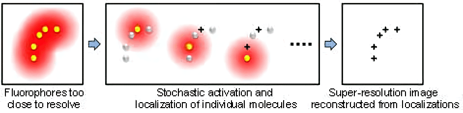
STORM (also named PALM) is a type of super-resolution optical microscopy technique based on stochastic switching of single-molecule fluorescence signal. In conventional fluorescence microscopy where all fluorophores in the sample are fluorescent, their diffraction limited images overlap, creating a smooth but blurred picture. STORM/PALM utilizes fluorescent probes that can switch between fluorescent and dark states so that in every snapshot, only a small, optically resolvable fraction of the fluorophores is detected. This enables determining their positions with high precision from the center positions of the fluorescent spots. With multiple snapshots of the sample, each capturing a random subset of the fluorophores, a final super-resolution image can be reconstructed from the accumulated positions. The scheme of STORM/PALM is illustrated below.

Mobirise


STORM has gained much functionality since its invention in 2006. Multicolor imaging can be achieved with photoswitchable fluorophores with either different emission wavelengths or different activation wavelengths. 3D imaging has been realized with various 3D single-particle localization methods, including astigmatic imaging, bi-plane imaing, PSF engineering and intereference. Live cell STORM has been demonstrated using either fluorescent proteins or organic fluorophores

Microtubules (green) and mitochondria (magenta) in BS-C-1 cells. Showing the conventional wide-field fluorescence image, the 2D STORM image, and a 50 nm thick z secion of from the 3D STORM image which resolves the hollow shape of the mitochondria outer membrane.

Mobirise

Useful Super-Resolution Microscopy Reviews

  • B. Huang, H. Babcock, X. Zhuang, “Breaking the diffraction barrier: super-resolution imaging of cells”, Cell, 143, 1047-1058 (2010) [link] [supp info] (The supporting information contains a detailed discussion of fluorescent probes for single-molecule-switching-based super-resolution microscopy methods).
  • S. Hell, "Microscopy and its focal switch", Nat. Methods, 6, 24-32 (2009) [link].
  • B. Huang, M. Bates, X. Zhuang, “Super resolution fluorescence microscopy”, Ann. Rev. Biochem., 17, 993-1016 (2009) [link].
  • S. Hell, "Far-field optical nanoscopy", Science, 316, 1153-1158 (2007) [link].
  • R. Heintzmann, M. G. L. Gustafsson, "Subdiffraction resolution in continuous samples", Nat. Photonics, 3, 362-364 (2009) [link].
  • D. Kamiyama B. Huang, "Development in the STORM", Developmental Cell, 23, 1103 (2012) [link].
  • D.M. Shcherbakova, P. Sengupta, J. Lippincott-Schwartz, V.V. Verkhusha, "Photocontrollable Fluorescent Proteins for Superresolution Imaging", Ann Rev Biophys, 43, 303-329 (2014) [link].
  • M. Fernandez-Suarez, A. Y. Ting, "Fluorescent probes for super-resolution imaging in living cells. Nat. Rev. Mol. Cell Biol., 9, 929-943 (2008) [link].

Huang Lab | UCSF Mission Bay Campus | Byers Hall 304 | 1700 4th St, MC 2532, San Francisco, CA 94143 | Fax: (415) 514-1028

Made with ‌

Landing Page Software